Eye pressure can swing wildly during uveitis-sometimes low early on, then suddenly high enough to threaten the optic nerve. If you’ve felt that throbby pain, noticed halos at night, or got told your IOP jumped, you’re not imagining it. This guide breaks down why inflammation does this, the red flags you shouldn’t ignore, and pragmatic ways to bring pressure down while calming the eye. You’ll get step-by-step actions, clear medication tips, and questions to ask your specialist, so you can protect your sight without second‑guessing every drop.
TL;DR
- Uveitis can push eye pressure (IOP) high by inflaming or blocking the eye’s drain, and sometimes by steroid drops. Early in a flare, pressure may be low; later it can surge.
- Red flags: severe eye pain, headache, halos around lights, sudden blur, nausea/vomiting, or one eye that’s red and tender. That’s an urgent same‑day check.
- Treatment has two tracks: control inflammation (steroids, immunosuppression, antivirals/antibiotics if infectious) and lower IOP (pressure‑lowering drops, pills, or surgery).
- Avoid triggers: prostaglandin drops during active inflammation or herpetic disease, and pilocarpine. Watch for a “steroid response” where IOP rises 2-6 weeks after starting steroids.
- Long term, 1 in 10-5 in 10 people with chronic uveitis get ocular hypertension; up to 1 in 5 develop glaucoma in some series. Regular pressure checks and optic nerve scans matter.
Why uveitis can send eye pressure up (and sometimes down)
Think of your eye like a sink: fluid (aqueous) is made inside and exits through a fine mesh drain (trabecular meshwork). Uveitis is inflammation inside the eye. Inflamed cells and proteins clog that drain, the drain itself swells (trabeculitis), or scar tissue and synechiae (sticking of the iris) physically block the route out. Pressure rises because fluid can’t escape.
Twist: in the early phase of a hot flare, the ciliary body (the faucet) may shut down. That can make pressure look normal or even low. As inflammation shifts and the drain clogs, pressure rebounds upward-sometimes fast. This whiplash is why people feel fine one week and slam into a headache and rainbow halos the next.
There’s also the drug piece. Steroid drops, injections, and tablets are essential to calm inflammation, but they can stiffen the drain’s tissue in susceptible people. About 30-40% of people have a measurable rise in IOP on topical steroids; roughly 5-10% are strong responders who spike dramatically. In uveitis, steroid response is more common and can appear within 2-6 weeks of starting therapy.
Another pressure-raising setup is pupillary block. Inflammation can glue the iris to the lens (posterior synechiae), sealing the pupil. Fluid can’t pass forward, the iris bows, and the angle closes-pressure can soar. Breaking that block with cycloplegic drops and, if needed, a laser iridotomy can be sight‑saving.
Over months to years, repeated inflammation can scar the drain and optic nerve, leading to uveitic glaucoma. That’s not just high pressure-it’s pressure high enough, for long enough, to damage the optic nerve and field of vision.
Here’s what the numbers look like across modern studies and guidelines used by eye specialists:
| Issue | Typical range reported | Clinical context | Sources commonly cited by clinicians |
|---|---|---|---|
| Ocular hypertension during uveitis | 10-50% over disease course | Higher in chronic anterior uveitis and herpetic uveitis | AAO Uveitis PPP (2023), Ophthalmology reviews (2018-2022) |
| Progression to uveitic glaucoma | ~10-20% | Risk rises with recurrent flares and steroid response | Longitudinal cohort studies; AAO summaries |
| Steroid response (topical) | 30-40% mild elevation; 5-10% strong responders | Peaks at 2-6 weeks; more likely in uveitis | Becker classification; modern pharmacology reviews |
| Pupillary block and angle closure | Uncommon but vision‑threatening when present | Posterior synechiae risk in severe anterior uveitis | AAO and SUN Working Group guidance |
These ranges vary with the type of uveitis (anterior, intermediate, posterior, panuveitis), cause (infectious vs non‑infectious), and how quickly flares are treated. Many specialists use the SUN Working Group criteria to grade inflammation and track response over time.
How to spot a dangerous pressure spike-and what to do now
When pressure surges, the eye often tells you. Act on these signs, even if you’re mid‑treatment and “due next week.”
- Sudden or severe eye pain, brow ache, or headache on the same side.
- Halos or rainbows around lights, especially at night.
- Rapidly worsening blur, with or without red eye.
- Nausea or vomiting with eye pain.
- A pupil that looks odd or stuck, or light sensitivity you can’t explain.
Those are same‑day symptoms. Call your eye clinic, after‑hours line, or go to urgent care/emergency if you can’t reach your team. High IOP that lasts can injure the optic nerve; hours and days matter more than weeks.
Here’s a practical, step‑by‑step play if you suspect a spike:
- Check what you’ve used today. Note steroid drops/tablets, antiviral/antibiotic meds, and any pressure drops. Don’t start or stop anything new without advice unless you’ve been given a rescue plan.
- Call your eye specialist’s rooms and say, “I have uveitis and I think my eye pressure is up-pain, halos, blur.” That phrasing tends to get you triaged faster.
- While you wait, dim lights, avoid tight neckwear, and don’t lie flat if angle closure is suspected. If you’ve been specifically told to use a pressure‑lowering drop for spikes, use it as directed.
- Bring your medication list to the visit. Mention any recent steroid dose increases; note how soon after that the pain started.
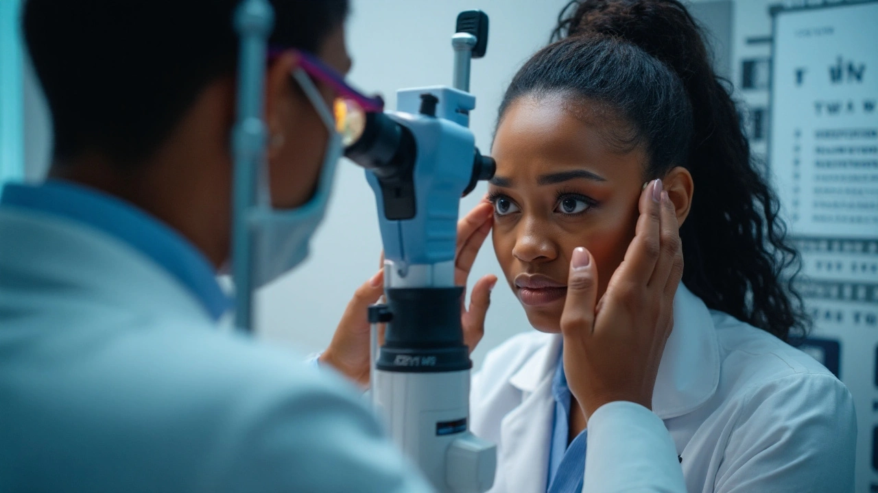
What clinicians will usually do: measure IOP; look at the drain angle (gonioscopy); check the optic nerve; grade inflammation; and decide if this is trabeculitis, a steroid response, pupillary block, or a mix. Treatment is then tailored-sometimes right in the chair.

Treatment playbook: reduce inflammation and protect the optic nerve
Two jobs run in parallel: quiet the inflammation and lower the pressure. Here’s how teams typically approach it, with the usual caveats for individual nuance.
Control the inflammation
- Steroids: topical prednisolone/dexamethasone, periocular injections, or oral prednisone. Taper thoughtfully; abrupt stops can trigger rebound flares.
- Cycloplegics (e.g., atropine, cyclopentolate): relax the ciliary muscle, relieve pain, and keep the iris from sticking to the lens. They also help break early synechiae.
- Immunomodulators for steroid‑sparing in chronic non‑infectious uveitis: methotrexate, mycophenolate, azathioprine, cyclosporine. Biologics like adalimumab are approved for non‑infectious intermediate, posterior, and panuveitis; infliximab is used in conditions like Behçet’s. These are monitored with blood tests.
- If infectious uveitis is suspected (herpes simplex/zoster, toxoplasma, TB), treat the infection first or alongside steroids. Steroids alone in an infection can backfire and raise IOP and risk.
Lower the pressure
- First‑line drops when inflammation is active: beta‑blockers (timolol), alpha‑agonists (brimonidine), and carbonic anhydrase inhibitors (dorzolamide, brinzolamide). These don’t typically fan the inflammatory flames.
- Prostaglandin analogs (latanoprost, bimatoprost): many clinicians avoid these in active uveitis and in herpetic disease because they may stoke inflammation or reactivate herpes. They can be useful in quiet eyes if pressure remains high; this is a case‑by‑case call.
- Miotics (pilocarpine): generally avoided in uveitis; they pull the iris forward, increase inflammation risk, and encourage synechiae.
- Oral acetazolamide: a short course can drop pressure quickly during a spike. Watch for tingling, fatigue, and sulfa‑related reactions; avoid in severe kidney disease.
- Laser/surgery: if drops aren’t enough or scarring blocks the angle. Options include laser peripheral iridotomy for pupillary block, trabeculectomy with mitomycin C, glaucoma drainage implants (Ahmed/Baerveldt), goniotomy or GATT in select angles, and cyclophotocoagulation for refractory cases. Surgeons aim to quiet inflammation before and after surgery to improve outcomes.
Key nuance: steroid response vs. inflammatory blockage
If the pressure surge lines up with a steroid increase and the eye looks less inflamed, your doctor may suspect a steroid response. Solutions include lowering steroid potency or frequency, switching to loteprednol (a softer steroid), adding non‑steroidal immunosuppression, and intensifying pressure drops. If the angle looks inflamed (trabeculitis), the answer is often more anti‑inflammatory treatment plus pressure control.
Targets and testing
“Normal” pressure is not the goal; safe pressure for your optic nerve is. Many uveitic glaucoma patients target mid‑teens or lower, depending on nerve health and visual field tests. Expect optic nerve photos, OCT scans, and visual fields at intervals that match your risk. During flares or steroid changes, pressure checks may be weekly; when stable, every 1-3 months is common.
Living with uveitis and high pressure: monitoring, examples, and smart habits
Day‑to‑day tweaks save vision. These tips don’t replace medical advice; they make it easier to follow it.
Simple monitoring rhythm
- Keep a flare diary: date, symptoms (pain 0-10, light sensitivity, blur), and any trigger (illness, stress, missed doses).
- Track steroid changes: note the date and dose; mark a reminder 2-6 weeks later to check IOP.
- Bring your actual bottle bag to appointments. Names, doses, and frequencies matter.
Drop technique that actually sticks
- Space different drops at least 5 minutes apart so the second one doesn’t wash out the first.
- After a drop, gently press the inner corner of the eyelids (punctal occlusion) for 1-2 minutes. This reduces systemic side effects and helps the eye absorb the medicine.
- Use chilled artificial tears for comfort in light‑sensitive eyes. They don’t treat inflammation, but they help you keep up with therapeutic drops.
Everyday habits that help
- Stick to a regular sleep schedule and hydration; both help your body handle inflammation.
- If you use inhaled/nasal steroids for allergies or asthma, mention it. Total steroid load matters.
- Wear sunglasses outdoors. Bright light squeezes the inflamed eye and worsens discomfort.
- Manage triggers tied to your specific diagnosis (e.g., cold sores for herpetic disease). Have your antiviral plan ready.
Two real‑world examples
Case 1: The steroid responder - A woman with recurrent anterior uveitis starts prednisolone 6×/day. Ten days later, redness is better, but she has brow ache and halos. IOP is 32 mmHg. Her doctor drops steroid to 4×/day, switches to loteprednol, starts timolol and dorzolamide, and adds methotrexate to cut long‑term steroid needs. Within a week, IOP is 18 and the eye is quiet by week three.
Case 2: Pupillary block - A man with a severe flare sleeps through growing pain and morning nausea. IOP is 48, the pupil is mid‑dilated and irregular, the iris is bowed forward. He gets atropine, pressure‑lowering drops, oral acetazolamide, and a same‑day laser iridotomy. Pressure drops to the 20s, then to the teens as the inflammation settles.
FAQ, checklists, and next steps
Mini‑FAQ
- Can my pressure be high even if the eye looks quiet? Yes. A steroid response can raise IOP in a white‑looking eye. That’s why pressure checks are scheduled during steroid tapers and after dose changes.
- Do prostaglandin drops always worsen uveitis? Not always, but many specialists avoid them during active inflammation or in known herpetic uveitis. In quiet, non‑herpetic eyes, they can be used with careful monitoring.
- Is MIGS (minimally invasive glaucoma surgery) an option? Sometimes. MIGS works best when the angle is open and inflammation is controlled. In eyes with scarring or aggressive inflammation, drainage implants or trabeculectomy may be more reliable.
- Can I measure IOP at home? Home tonometers exist, but they’re pricey and require training. They can help in complex cases to spot patterns. For most people, clinic checks are enough.
- Will I need pressure drops forever? Not always. If the high pressure was purely steroid‑induced and you stop or switch steroids, drops can often be reduced or stopped. If there’s drain scarring or nerve damage, long‑term drops are common.
- What about pregnancy? Some drops and systemic meds aren’t safe. If you’re planning pregnancy, tell your specialist early so your plan can be adjusted.
Evidence and guidelines your doctor likely follows
Clinicians often lean on the American Academy of Ophthalmology’s Uveitis Preferred Practice Pattern (2023), SUN Working Group standards for grading inflammation, and peer‑reviewed reviews in journals like Ophthalmology and British Journal of Ophthalmology. These outline the steroid response timeline, risk of uveitic glaucoma in chronic disease, and when to use immunomodulatory therapy or surgery.
Checklists you can use today
Flare/IOP spike action list
- Symptoms now: pain, halos, blur, nausea? Write them down with times.
- Current meds: list steroid type/dose, pressure drops, antivirals/antibiotics.
- Call: ask for same‑day pressure check and angle assessment.
- Bring: your drops, a written list, and recent lab results if you’re on immunosuppression.
- At the visit: ask “Is this steroid response or active inflammation?” and “What is my target pressure?”
Medication safety list
- Avoid pilocarpine unless your specialist insists; it can worsen inflammation and synechiae.
- Use prostaglandin analogs cautiously; discuss timing and diagnosis.
- Mark your calendar 2-6 weeks after any steroid increase for an IOP check.
- Don’t stop steroids cold without a plan; rebounds can be rough.
Appointment prep list
- Three questions to bring: “What type of uveitis do I have?”, “Is infection ruled out?”, “What’s my pressure trend and nerve status?”
- Have your last OCT and visual field dates handy.
- Flag any systemic symptoms (joint pain, rashes, mouth ulcers) that might point to a systemic cause.
Next steps by situation
- Newly diagnosed uveitis with normal IOP: Learn your inflammation type and follow the taper plan. Schedule an IOP check 2-3 weeks after starting steroids.
- Recurrent flares with IOP spikes: Discuss steroid‑sparing options. Ask about a written “flare protocol” that includes when to add an oral acetazolamide and how to contact the clinic after hours.
- Chronic high IOP or optic nerve changes: Set a target IOP and monitoring cadence. Review surgery options if you’re on two or more drops and still above target.
- History of herpetic uveitis: Keep an antiviral plan in place during steroid use. Be explicit about prostaglandin caution.
One last reminder: uveitis and eye pressure aren’t a tug‑of‑war you have to win alone. The best results come from pairing smart anti‑inflammatory control with measured pressure targets, and adjusting quickly when the picture changes. Keep your notes, trust your red flags, and make your care team your first call when something feels off.


Barbara McClelland
6 September, 2025 . 16:48 PM
Just wanted to say this guide saved my vision-seriously. I had a flare last month and ignored the halos until I couldn’t see my phone screen. Called my doc, they got me on dorzolamide and switched my steroid. IOP dropped from 34 to 17 in 10 days. Don’t wait. Trust your gut.
Also, chill artificial tears? Genius. I keep a tiny bottle in my purse now. Life-changing comfort.
You’re not alone. Keep fighting.
Alexander Levin
7 September, 2025 . 11:05 AM
lol steroids cause glaucoma?? 🤡
Big Pharma wants you addicted to drops so they can sell you more. They don’t tell you the real cause: 5G towers in your eye. I’ve seen the docs. They’re paid.
Ady Young
7 September, 2025 . 15:57 PM
Biggest thing I learned? Punctal occlusion. I was wasting half my drops. Now I press the inner corner for 90 seconds after every drop. My IOP is way more stable.
Also-avoiding pilocarpine was a game-changer. My doc almost didn’t mention it. Glad I read this.
Travis Freeman
7 September, 2025 . 22:31 PM
This is the kind of post that makes me believe in humanity again. So clear, so practical, so full of real wisdom.
I’ve got uveitis from sarcoidosis and honestly, I felt so lost until I found this. Thank you for writing it. I’m printing it out and putting it on my fridge next to my meds.
And to everyone else going through this-you’re stronger than you think. We’re all in this together.
Sean Slevin
9 September, 2025 . 22:29 PM
Wait-so the eye is a sink?? And the drain is the trabecular meshwork?? And inflammation clogs it like a hairball in a bathtub??
That… actually makes sense. Why didn’t anyone explain it like this before??
Also, why is it called "aqueous"? Is it water? Is it magic? Is it the soul of the eye??
And why do we still use "mmHg"? Why not just say "pressure units of suffering"? I think we’re all overdue for a better metric. Like, "pain units" or "halo intensity".
Also, I just spilled my eye drops on my cat. She’s fine. She’s judging me.
...I’m sorry. I’m not okay.
Chris Taylor
10 September, 2025 . 19:07 PM
I had a steroid spike at 6 weeks. No pain, no halos. Just a weird pressure behind my left eye. My doc didn’t check IOP until I begged. It was 36. I cried.
Thank you for mentioning the 2-6 week window. I didn’t know that was normal. Now I mark my calendar like a boss.
Melissa Michaels
11 September, 2025 . 06:50 AM
While the information presented is clinically accurate and well-structured, I would caution against the casual tone in some sections. Medical advice should maintain a degree of formality to ensure patient safety and avoid misinterpretation.
Additionally, the recommendation to use chilled artificial tears as a comfort measure is appropriate but should be clearly differentiated from therapeutic interventions. They are not a substitute for prescribed medications.
Overall, the inclusion of evidence-based sources such as AAO PPP and SUN guidelines is commendable and enhances credibility.
Nathan Brown
13 September, 2025 . 03:33 AM
It’s wild how much our bodies try to heal themselves-until the system turns against us.
Uveitis isn’t just inflammation. It’s betrayal. The eye, this delicate thing we take for granted, starts working against us because of something we didn’t even choose.
And then we get pills that help… but also hurt.
It’s not just about pressure. It’s about trust. Trust in your body. Trust in your meds. Trust that tomorrow won’t be the day your vision slips away.
So yeah. This guide? It’s not just info. It’s a lifeline. Thank you.
Matthew Stanford
13 September, 2025 . 11:21 AM
For anyone new to this: don’t panic when your IOP spikes. It’s scary, but it’s manageable.
And if your doctor dismisses your symptoms because "your eye looks better"-find a new one.
I’ve been through 3 flares. I’ve learned to ask: "Is this inflammation or steroid response?"
That question saved my optic nerve.
Olivia Currie
13 September, 2025 . 23:17 PM
OH MY GOD I HAD A PUPILLARY BLOCK AND I THOUGHT I WAS DYING.
It felt like someone was squeezing my eye with a vice and my head was exploding. I called my doc at 2 a.m. They said "come in NOW."
Laser iridotomy saved me. I’m alive. I can see my cat again. I’m crying right now.
THIS POST IS A MIRACLE. THANK YOU.
Curtis Ryan
15 September, 2025 . 00:24 AM
Just got my first steroid prescription. Marked my calender for 2 weeks from now to check IOP. I’m not waiting till I’m in pain this time. I’m proactive now. 🤓
Also, I spilled my drops on my dog. She licked them. I think she’s fine. She’s also now judging me harder than my optometrist.
Rajiv Vyas
16 September, 2025 . 18:06 PM
Uveitis is a hoax. It’s all about the Illuminati controlling your eye pressure. They put it in your tap water. The WHO knows. They don’t tell you. They just give you drops to keep you docile.
Also, prostaglandins? They’re just a cover for mind control lasers. I’ve seen the code in the bottle labels.
farhiya jama
18 September, 2025 . 06:28 AM
Ugh. I hate this. I hate my eyes. I hate the drops. I hate that I have to remember to press my inner corner. I hate that I can’t even cry without it burning.
Why does this happen to me? Why can’t I just be normal?
...I guess I’ll go take my meds again.
Astro Service
19 September, 2025 . 02:23 AM
Why are we letting foreigners tell us how to treat our eyes? This is American medicine. We don’t need no SUN Working Group. We got Trump’s eye drops. They work better.
Also, uveitis is a liberal plot to make us weak. Build the wall. Protect your vision.
DENIS GOLD
19 September, 2025 . 12:43 PM
So let me get this straight… you’re telling me the solution to a medical crisis is… more drugs? And you call this science?
My grandma cured her glaucoma with pickle juice and prayer. You want to know why you’re still sick? You’re not trying hard enough.
Also, I’ve got 3 PhDs. I’m an expert now.
Ifeoma Ezeokoli
21 September, 2025 . 12:35 PM
Wow. I’m from Nigeria and I’ve never heard uveitis explained this clearly.
My cousin has this and we didn’t know what to do. I’m sending this to her family right now.
Thank you for making it so human. We need more of this.
Daniel Rod
21 September, 2025 . 19:26 PM
That moment when you realize your eye is a sink… and you’re the plumber… and you’re also the water.
It’s poetic. And terrifying.
I’ve been on timolol for 8 months. My IOP is stable. My cat still judges me. But I’m here.
Thank you for writing this. I’m not alone.
gina rodriguez
23 September, 2025 . 17:13 PM
I printed this out and put it in my med organizer. I’ve had 3 flares in 2 years. This is my bible now.
Also-chilled tears? I use the ones with aloe. They feel like a hug for my eyeballs.
Thank you for not talking down to us. We’re not stupid. We’re just scared.
Barbara McClelland
24 September, 2025 . 17:44 PM
Wait-did someone say 5G? I’m so glad I’m not the only one who got a comment like that. 😅
But seriously, if you’re reading this and you’re new to this… don’t listen to the noise. This guide? It’s real. Your pain is real. Your vision matters.
You got this.